Among the diseases are sinusitis frontal sinusitis etmoiditois and.
Floor of left maxillary sinus.
When the area shows up white or gray it is called opaque or opacification of the sinus.
In some cases the floor can be perforated by the.
This is the reason for the late detection of diseases.
The cyst of the left as well as the right maxillary sinus does not manifest itself in symptomatology for a long time and is only detected with radiography or tomography.
The maxillary sinus drains into the nose through a hole called the ostia when the ostia becomes clogged sinusitis can occur.
Because the symptoms manifest themselves in a serious complication of the disease.
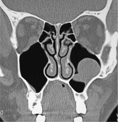
The floor is formed by the alveolar process and if the sinus is of an average size is on a level with the floor of the nose.
The maxillary sinus is a dual paranasal sinuses which are located throughout the maxilla.
When a ct scan is taken of the head the sinuses should show up black since they are cavities.
Causes the sinuses to be swollen.
Within the maxillary sinus which lies beneath the cheek bone on each side are mucous glands a blockage in the mucous duct can cause the gland to enlarge which can lead to the formation of a dome shaped maxillary mucous retention cyst the cyst does not usually cause any symptoms and does not damage expand or thin the wall of the sinus.
My brother s cemri shows diffuse mucosal thickening are seen in both ethmoidal both maxillary sinuses more on left side answered by dr.
Cyst of left and right maxillary sinus.
These cavities are called sinuses and they are located in the maxilla or upper jaw cysts are closed pocket like formations of tissue and are filled with liquid air or semi solid material.
Maxillary sinus floor augmentation also termed sinus lift sinus graft sinus augmentation or sinus procedure is a surgical procedure which aims to increase the amount of bone in the posterior maxilla upper jaw bone in the area of the premolar and molar teeth by lifting the lower schneiderian membrane sinus membrane and placing a bone graft.
The maxillary sinus is the cavity behind your cheeks very close to your nose 1.
Without an exam i.
Inside has a mucous film that almost does not feel the elements due to the lack of nerve and blood vessels.
Of the symptoms can be identified random sudden single discharge from one side of the nose.
If the sinus is large it reaches below this level.
Often their formation is due to chronic diseases.
As there is no normal tissues regeneration and the excretory ducts patency of the mucous glands is not restored.
The ostia of the maxillary sinus often clog because the ostia are.
Maxillary sinus retention cysts are most often the result of inflammatory changes in the mucous membranes.