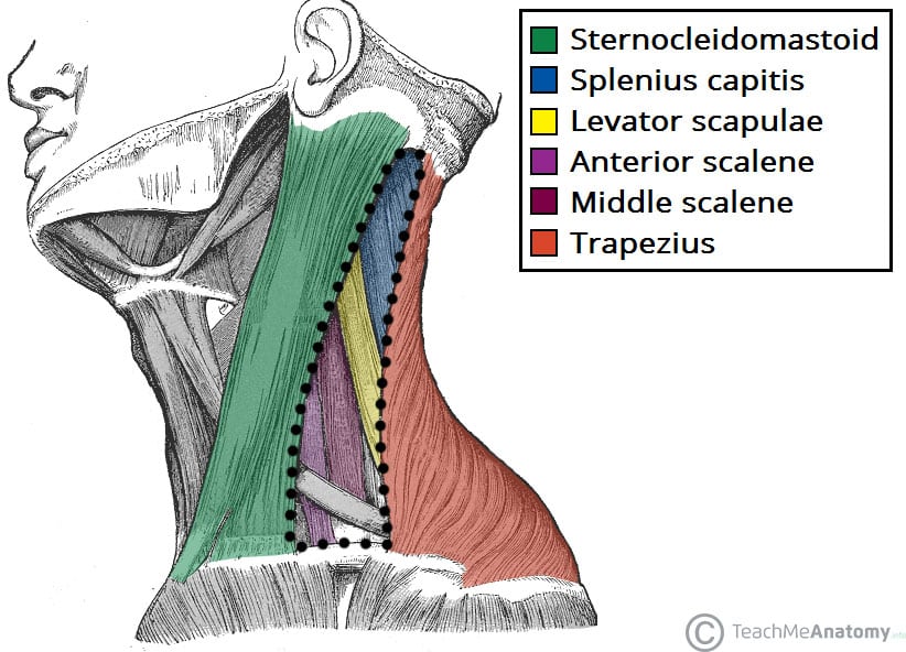
Floor Of Lateral Cervical Region
Posterior Triangle Of The Neck Subdivisions Teachmeanatomy
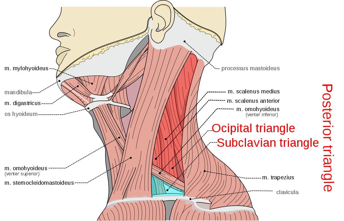
Posterior Triangle Of The Neck Wikipedia

Lateral Cervical Region Flashcards Quizlet

11 6 Axial Muscles Are Muscles Of The Head And Neck Vertebral Column Trunk And Pelvic Floor

Image Result For Anatomy Of Jaw And Ear Dental Anatomy Human Anatomy And Physiology Anatomy And Physiology

Gross Anatomy Of Neck Spencer Flashcards Quizlet

Union of the.
Floor of lateral cervical region.

Opening Of The Mandible Anatomy Retrodiscal Tissue Bilaminar Zone Retrodiscal Tissue Bilaminar Zone Expanded Flo In 2020 Anatomy Medical Knowledge Body Anatomy

Floor Of Fourth Ventricle Note Both Vagal Hypoglossal Triangle Facial Nerve Vagus Nerve Brain Anatomy

Musculature Of Pharynx Much Of The Framework Of The Lateral And Posterior Walls Of The Pharynx Is Formed By An In 2020 Plexus Products Medical School Studying Muscle

Ligaments Of The Pelvic Region Beckenboden Anatomie Gesundheit Und Wellness
Source : pinterest.com
